


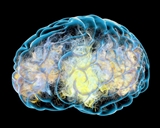
What Can Molecular Imaging Offer for the Differential Diagnosis of Parkinsonism?
 Date: May 2020
Prepared by SIC Member: Ece Bayram, MD, PhD
Authors: Cecilia Peralta, MD; David Eidelberg, MD
Editor: Un Jung Kang, MD
Parkinson’s disease and atypical Parkinsonian disorders may present with similar symptoms despite different underlying pathologies. Given that pathological processes associated with these disorders are yet to be fully clarified to enable discovery of effective treatments, the correct identification of patients is essential to aid in future research on pathogenesis, progression and treatment of these disorders. Despite the good sensitivity and specificity of the updated diagnostic criteria of Parkinsonian disorders, biomarkers are crucial to reliably differentiate these disorders. Molecular imaging is one of the biomarkers that can be used for differential diagnosis; as it allows visualization and quantification of cellular and subcellular processes. New advances in radionuclide tracers, which allow visualization of different chemical processes increase the value of this imaging technique for differential diagnosis of Parkinsonian disorders with different underlying molecular changes. To get a sense of molecular imaging’s current potential for utilization in the clinical setting and the barriers limiting the use, we asked two experts to share their opinions with us.
View definitions of terms in this article
|
1- Are there any molecular imaging techniques helpful for the differential diagnosis of parkinsonism currently?
Dr. Peralta: Yes, the most accessible approach is the use of a marker for the dopamine transporter (DAT). DaTscan is approved by FDA for routine clinical use in the differential diagnosis of Parkinson’s disease and Essential Tremor. This technology has shown 95% sensitivity and 93% specificity for the differential diagnosis of clinically diagnosed Parkinson’s disease and Essential Tremor. Moreover, it may also help to differentiate other causes of parkinsonism, such as those due to neuroleptic treatment, vascular or psychogenic parkinsonism. However, they are unable to differentiate Parkinson’s disease from other neurodegenerative parkinsonisms such as Multiple System Atrophy and Progressive Supranuclear Palsy and should not be used for this purpose. Presynaptic dopaminergic terminal scans may be helpful in conjunction with postsynaptic scans (such as raclopride, fallypride, or iodobenzamide SPECT among others) in the differential diagnosis of Parkinson’s disease and atypical Parkinsonian disorders. Dopamine receptor availability is typically decreased in Multiple System Atrophy and Progressive Supranuclear Palsy as opposed to Parkinson’s disease, where D2 receptors are normal to increased. However, receptor imaging compounds are not approved for clinical use by the FDA and most clinical centers do not perform this combination.
Another molecular imaging technique useful for differential diagnosis is the study of cerebral glucose metabolism with FDG-PET, which enables the quantification of network abnormalities expressed in a pattern-specific manner. FDG-PET studies have demonstrated to be superior to D2/D3 SPECT in differentiating Parkinson’s disease from atypical Parkinsonian disorders and the more recent diagnostic criteria of the Movement Disorder Society support the use of FDG-PET for discriminating Parkinson’s disease from atypical Parkinsonian disorders. Molecular imaging targeting alpha-synuclein holds great interest and new PET tracers are under investigation.
Dr. Eidelberg: Regarding the new tau-PET ligands, while valuable as a marker for Alzheimer’s disease, the story with these ligands in Parkinson’s disease and atypical Parkinsonian disorders is yet to be determined. The results thus far are encouraging for tauopathy syndromes -- particularly Progressive Supranuclear Palsy and perhaps also Corticobasal Degeneration. The meaning of the low-level changes seen in synucleinopathies such as Parkinson’s disease, Dementia with Lewy bodies or Multiple System Atrophy is less clear. In any event, formal studies are needed to assess the diagnostic accuracy of tau-binding ligands -- either alone or in conjunction with other tracers -- in individual patients with Parkinsonian disorders.
2- What are we lacking in the current molecular imaging approaches and what is required to develop reliable imaging markers?
Dr. Eidelberg: In a nutshell the major limitations for the use of current molecular imaging techniques in the clinical setting are:
- Availability of PET instruments for clinical use: Relevant tracers such as FDG-PET are commercially available and others are currently being developed for commercial distribution.
- Data processing/informatics: Automated diagnostic algorithms for FDG-PET are close to maturity and should be ready to be submitted for FDA approval this year. This essential first step followed by the development of "bulletproof" software for accurate differential diagnosis will take a few years. Industrial involvement may accelerate this process substantially.
Dr. Peralta: Despite these shortcomings, molecular imaging represents the best opportunities to understand the pathogenesis in parkinsonism. New technologies are emerging, such as the use of hybrid PET/MRI to incorporate the benefits that come with MRI to improve the spatial and temporal resolution. Artificial intelligence-based techniques and machine learning using automated diagnostic algorithms are in research to improve the objective analysis of different imaging modalities as well as the use of MRI atlas for automated segmentation in the differential diagnosis of Parkinson’s disease and atypical Parkinsonian disorders.
3- Do you think one imaging method will be enough for the differential diagnosis or should it be used in combination with other techniques?
Dr. Eidelberg: I think the ideal is a combination of dopaminergic imaging (ideally with F-DOPA or FP-CIT PET, which have greater signal-to-noise than DaTscan and related SPECT techniques) to document the presence of a presynaptic dopamine deficit -- and metabolic imaging (ideally with FDG-PET, although resting state-fMRI may ultimately prove suitable for this purpose) in conjunction with a pattern-based decision algorithm. The resting state-fMRI method was developed as an alternative to FDG-PET for probabilistic pattern-based diagnosis -- but more widely available and not requiring radiotracer injection. As a marker of functional network activity over the whole brain, resting state-fMRI and FDG-PET methods nicely complement localized neurochemical assays of in vivo nigrostriatal dopaminergic dysfunction (e g., F-DOPA/FP-CIT PET, DaTscan), amyloid/tau binding PET, etc).
Dr. Peralta: Based on all the advances in imaging methods that have been very instrumental in contributing to the understanding of the complex interactions between proteinopathies and network abnormalities, I consider that a combination of multi-modalities using PET and MRI and automated algorithms will substantially increase the accuracy of differential diagnosis. As we discussed before, presynaptic dopaminergic imaging (DATscan-F-DOPA PET) is effective to differentiate degenerative from non-degenerative parkinsonism and the combination of presynaptic dopaminergic imaging plus FDG-PET are useful to differentiate between Parkinson’s disease and atypical Parkinsonian disorders. Recent studies using MRI protocols including different markers of neurodegeneration--such as dorsolateral nigral hyperintensity and atrophy plus iron deposition, OR neuromelanin-sensitive images, dorsolateral nigral hyperintensity, diffusion tensor imaging and iron deposition--showed that the diagnostic accuracy in the differentiation of atypical Parkinsonian disorders from Parkinson’s disease increased by using a combination of these methodologies.
Conclusions:
The complexity of the brain and neurodegenerative diseases makes it difficult to find a biomarker that might serve them all, thus there is no blanket imaging technique, but instead many that serve specific functions in differentiating diagnoses. New ligands still need to be validated for clinical use with future research, but currently available molecular imaging techniques (DaTSCAN, FDG-PET) can indeed be useful for differential diagnosis. The PET and SPECT instruments are unfortunately not widely available, limiting the use. There is also a need for objective analysis methods to make the best of the available and upcoming molecular imaging approaches. Using multi-modal imaging by combining different molecular imaging methods and functional MRI can help increase the diagnostic accuracy and help us understand the pathogenesis of these complex diseases better. Overall, there is still a way to go to incorporate the routine use of molecular imaging at the clinical setting, but there is no denying that it is a strong biomarker candidate that is desperately needed.
References
- Eidelberg D, Moeller JR, Dhawan V, Spetsieris P, Takikawa S, Ishikawa T, Chaly T, Robeson W, Margouleff D, Przedborski S, et al. The metabolic topography of parkinsonism. J Cereb Blood Flow Metab. 1994 Sep;14(5):783-801.
- Antonini A, Leenders KL, Vontobel P, Maguire RP, Missimer J, Psylla M, Günther I. Complementary PET studies of striatal neuronal function in the differential diagnosis between multiple system atrophy and Parkinson's disease. Brain. 1997 Dec;120 ( Pt 12):2187-95.
- Benamer HTS, Patterson J, Grosset DG, Booij J, De Bruin K, Van Royen E, Speelman JD, Horstink MHIM, Sips HJWA, Dierckx RA, Versijpt J, Decoo D, Van Der Linden C, Hadley DM, Doder M, Lees AJ, Costa DC, Gacinovic S, Oertel WH, Pogarell O, Hoeffken H, Joseph K, Tatsch K, Schwarz J, Ries V. Accurate differentiation of parkinsonism and essential tremor using visual assessment of [123I]-FP-CIT SPECT imaging: The [123I]-FP-CIT study group. Mov. Disord. 2000; 15:503–510.
- Tang CC, Poston KL, Eckert T, Feigin A, Frucht S, Gudesblatt M, Dhawan V, Lesser M, Vonsattel JP, Fahn S, Eidelberg D. Differential diagnosis of parkinsonism: a metabolic imaging study using pattern analysis. Lancet Neurol. 2010; 9:149–158.
- Hellwig S, Amtage F, Kreft A, Buchert R, Winz OH, Vach W, Spehl TS, Rijntjes M, Hellwig B, Weiller C, Winkler C, Weber WA, Tüscher O, Meyer PT. [¹⁸F]FDG-PET is superior to [¹²³I]IBZM-SPECT for the differential diagnosis of parkinsonism. Neurology. 2012 Sep 25;79(13):1314-22.
- Vladimir Kepe, PhD,a Yvette Bordelon, MD, PhD,b Adam Boxer, MD, PhD,e Sung-Cheng Huang, DSc,a Jie Liu, PhD,a Frederick C. Thiede, BSc,b John C. Mazziotta, MD, PhD,b Mario F. Mendez, MD, PhD,b,c,f Natacha Donoghue, MA,c,d Gary W. Small, MD,c and Jorge R. Barrio, PhD. PET Imaging of Neuropathology in Tauopathies: Progressive Supranuclear Palsy. J Alzheimers Dis. 2013 Jan 1; 36(1): 145–153.
- Tripathi M, Tang CC, Feigin A, De Lucia I, Nazem A, Dhawan V, Eidelberg D. Automated Differential Diagnosis of Early Parkinsonism Using Metabolic Brain Networks: A Validation Study. J. Nucl. Med. 2016;57:60–6.
- Strafella AP, Bohnen NI, Perlmutter JS, Eidelberg D, Pavese N, Van Eimeren T, Piccini P, Politis M, Thobois S, Ceravolo R, Higuchi M,Kaasinen V, Masellis M, Peralta MC, Obeso I, Pineda-Pardo JÁ, Cilia R, Ballanger B, Niethammer M, Stoessl JA; IPMDS-Neuroimaging Study Group. Molecular imaging to track Parkinson's disease and atypical parkinsonisms: New imagingfrontiers. Mov Disord. 2017 Feb;32(2):181-192
- Hammes J, Drzezga A, van Eimeren T. The Role of Tau Imaging in Parkinsonian Disorders. Curr. Neurol. Neurosci. Rep. 2018;18:86.
- Schindlbeck KA, Eidelberg D. Network imaging biomarkers: insights and clinical applications in Parkinson’s disease. Lancet Neurol. 2018;17:629–640.
- Liu ZY, Liu FT, Zuo CT, Koprich JB, Wang J. Update on Molecular Imaging in Parkinson's Disease. Neurosci Bull. 2018; 34(2):330-340
- Pyatigorskaya N, Magnin B, Mongin M, Yahia-Cherif L, Valabregue R, Arnaldi D, Ewenczyk C, Poupon C, Vidailhet M, Lehéricy S. Comparative Study of MRI Biomarkers in the Substantia Nigra to Discriminate Idiopathic Parkinson Disease. AJNR Am J Neuroradiol. 2018 Aug;39(8):1460-1467
- Calloni SF, Conte G, Sbaraini S, Cilia R, Contarino VE, Avignone S, Sacilotto G, Pezzoli G, Triulzi FM, Scola E. Multiparametric MR imaging of Parkinsonisms at 3 tesla: Its role in the differentiation of idiopathic Parkinson's disease versus atypical Parkinsonian disorders. Eur J Radiol. 2018 Dec;109:95-100.
- Krismer F, Seppi K, Göbel G, Steiger R, Zucal I, Boesch S, Gizewski ER, Wenning GK, Poewe W, Scherfler C. Morphometric MRI profiles of multiple system atrophy variants and implications for differential diagnosis. Mov Disord. 2019 Jul;34(7):1041-1048.