




What is it? A young girl with subacute jerky movements



Dr. Hugo Morales Briceno: [00:00:00] Welcome to What Is It?, a clinical case discussion podcast series. In each episode, we dissect a movement disorder case step by step, starting with the history and examination and moving through phenomenology, neuroimaging, laboratory findings, and a final diagnosis. The focus here isn't on getting the answer quickly.
It's about making expert reasoning visible, how hypotheses are formed, revised, and sometimes discarded as new information emerges. I'm your host, Hugo Morales, and in this episode, I'm discussing a case with Dr. Kathryn Yang, pediatric neurologist and movement disorder specialist from Boston Children's Hospital.
View complete transcript
Welcome, Kathryn.
Dr. Kathryn Yang: Thank you for having me, Hugo
Dr. Hugo Morales Briceno: It's a pleasure. Now to prepare our audience, I would like t- to start by reading the [00:01:00] clinical history, and then I would like to get your thoughts on the presentations before moving into the examination. Now, this is a previously normal 15-year-old girl who presented with a gradual symptom onset over a couple of weeks.
The symptoms are, she described, were jerky upper limb movements that were present at rest and also with action. Over a period of one month, her symptoms all have progressed in this case, and also her gait becomes unsteady. On specific questioning, her symptoms have developed after a febrile illness.
However, we don't have details about the severity of the such a febrile illness. Her medical history was unremarkable, and she was not taking any medications. Now, if we start with this, Kathryn, so in after hearing the clinical presentation, what comes to your mind regarding the possible [00:02:00] findings?
Dr. Kathryn Yang: Yeah, I think it's a great question, and I think starting off pretty broad. Just thinking about the stem in general, it's important to highlight that this is a previously normal 15-year-old girl. I think when we start later on building a differential, it helps categorize in buckets what you're thinking about and your underlying etiology.
The gender might be something that we think about specifically when it comes to this case as well. So keeping that in mind. The cadence of the symptoms hint at sort of a subacute picture. So again, something to think about when we think about patterns for certain syndromes or etiology.
And I think the important part will come when we think about the exam. So right now, the description of her movements is this jerky upper limb movement at rest and with action, and it's rather re-relatively nonspecific. It hints at potentially a hyperkinetic, because we're thinking about movement disorder, hyperkinetic movement disorder.
But under that umbrella term, there's a lot of different phenomenologies [00:03:00] that we can point to. So one key question later on is defining what the leading phenomenology is. And this sort of history of progression and involving her gait as well will be something to keep in mind, along with the inciting event of a febrile illness, given that infection and post-infection sort of movement disorders can be quite common and there's various things on our list of differentials. But overall, it still leaves a lot of open questions, and I think ties us nicely into thinking about the upcoming video and also the investigations that we'll be looking into.
Dr. Hugo Morales Briceno: I agree. The history is telling you something happened subacutely, and then the clinical find is gonna be very helpful to understand what's happening. Now, I'm gonna give you the clinic examination, and then I'll ask you to describe the phenomenology of the video that I have shown you.
Now, on examination, she was alert, oriented, but the Mini-Mental State Exam scored [00:04:00] 18, losing points on attention. But her language and speech was preserved. Motor strength and reflexes were normal, as well as sensation. There was no upper limb ataxia by the description, but-- and the main findings was that this patient had these generalized jerky movements.
Now on the video what are you seeing? If you could tell the audience what you're looking at and what are you thinking is happening?
Dr. Kathryn Yang: For sure. So I'm just looking at the video as we speak to walk us through, but this is a teenage girl. She's lying in a hospital bed. She's awake and she's following command appropriately. But you can see from the start that there's a couple signs that are notable.
So the first part is in her arms. So I'm guessing the examiner is asking her to hold her arms up in a posture. And when she does that, there's these continuous, what look like jerky movements in bilateral arms. They [00:05:00] don't disappear at any point of the exam, but she's also holding her arm up the entire exam as well.
So with posture, there's this elicited hyperkinetic movement. And if I were to see this the leading phenomenology that I would call this is myoclonus. I think you get there pretty quickly just from the bedside sort of exam because the movements are jerky. They occur what was described at both at rest and with action and they're generalized.
So at a later point in the exam, you can see that they also involve her feet, so her lower limbs are also affected. And I think a sort of a pearl for learners is that this sort of argues against, tremor, for example, which is typically really rhythmic and oscillatory around a joint.
This is arrhythmic. It's very sudden. It's shock-like. And so I think myoclonus would be top of the mind. The other clinical finding that stands out is in her eye movements.
So when you look at her [00:06:00] in the bed, she's sitting there, but she has these what look like very chaotic, rapid, multidirectional eye movements.
And that sort of description sometimes clues us into something called opsoclonus. Opsoclonus is typically involuntary it's conjugate, it's multidirectional and it occurs back to back without a normal intersaccadic interface which helps you distinguish this from another type of eye movement that, that can sometimes look a little bit similar called ocular flutter.
But that tends to be purely horizontal. And it also distinguishes this from nystagmus, whi-which is which has a slow phase followed by a fast corrective phase. So these eye movements are neither slow phase nor resting interval. It's very chaotic. It's in every direction, and it's sustained throughout the entire video.
Dr. Hugo Morales Briceno: Yeah. But l- the description that you've given us is she looks very syndromic with a combination of generalized myoclonus and the opsoclonus, which is a very distinctive finding. And I guess [00:07:00] as the early learners of neurology and resident fellows, they haven't seen a myoclonus it's easy to c-confuse it by other things for, nystagmus is more had the quick phases and slow phases, has a varied direction, but this description that you've given us is very clear of opsoclonus, so there's no doubt that this is the syndrome.
Now, in your practice how would you approach them just in, in the background that you mentioned us to? She's subacute, there's some febrile illness, then you find opsoclonus myoclonus. How would you like to investigate this patient?
Dr. Kathryn Yang: So I think before thinking about additional tests, like you said there's some sort of syndromic description here that fits something called-- So when I think-- when we think about opsoclonus myoclonus some gait ataxia, and potentially some mild encephalopathy with the mini-mental assessment in a previously well adolescent, we think of [00:08:00] something called opsoclonus myoclonus ataxia syndrome, or OMAS.
There's an older nickname for this. It used to be called dancing eyes, dancing feet. And I think it's helpful to, to frame it in this way. So from a diagnostic perspective, if three of the four features are present, so opsoclonus, ataxia or myoclonus, behavioral change or sleep disturbances, and later on we'll discuss underlying etiologies like neuroblastoma.
If three of the four features are present, then this diagnosis can be made. And it's more of a description of the syndrome. I think once you've made that identification we then have to think about, what is the underlying trigger for this? What could be causing these symptoms? And so I think that sort of then leads you into dedicated testing.
I think if we were to approach this patient just at the bedside, one question that you'd want to answer is, sometimes myoclonus can be epileptic in nature, so differentiating between myoclonus that is [00:09:00] coming from the cortex or cortical myoclonus versus subcortical and beyond because that would also change your approach but also your treatment modalities.
So that might be the first thing that you would think about. But I would start with that and then also I think testing for myoclonus, specifically doing something called an EMG as well, because sometimes the EMG morphology can give you some information.
Dr. Hugo Morales Briceno: I have some results for you. So the patient had an electroencephalogram that was reported to be normal. She also had a polymyography that showed this high-frequency burst less than one hundred milliseconds. And the EEG, EMG jerk locked, there was no correlation or cortical correlation to the jerks meaning that they're not coming from the cortex, at least.
The other investigations are relevant is that her brain MRI was normal and systemic investigations were also normal. There was no [00:10:00] descriptions of liver dysfunction, renal problems. And the CSF showed two cells protein of forty-eight is slightly elevated in normal glucose.
And she also had investigations with a CT abdominal and chest scan that were reported normal. With this in mind, with these results how would you like to move forward into other investigations?
Dr. Kathryn Yang: Yeah. So I think to summarize the investigations that have been performed so far. So I think what the EEG and the EEG EMG tells us is that these are not seizures. So that's something that we've excluded. The EEG is normal and there's no actual correlation. It also, definitively tells you that there's no correlation between the EMG and the EEG itself with these jerk-like movements or the myoclonus.
The EMG morphology tells you a little bit as well about where potentially things could be localized. Cortical myoclonus [00:11:00] typically produces very brief bursts somewhere in the 10 to 50-millisecond range. What we're seeing in this presentation, in this investigation is polyphasic bursts under about 100 milliseconds, which is a little bit longer, but fits much better with a subcortical generated pattern.
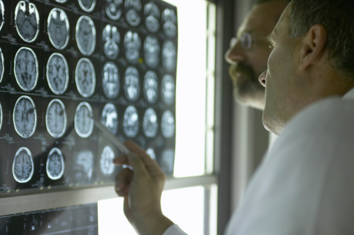
So I think that's something that we can pull away from this. The brain MRI is reassuring in that it doesn't show any structural lesions. So oftentimes we think about changes in the basal ganglia or inflammatory pictures or if you think about the ataxia changes to the cerebellum, that might be explaining some of these symptoms, and the brain MRI is unremarkable.
As you mentioned, her systemic investigations haven't shown anything, and her CSF, despite showing the two cells, really is relatively bland. So again it doesn't show a true lymphocytic pleocytosis and the CT abdo/chest as a first point is unremarkable. So I think when we think about broadening our tests, [00:12:00] especially in this clinical scenario where we're assuming that this is an OMAS picture.
I think organizing the expanded screening into sort of three buckets. So I think important for OMAS tumor screening would be one bucket. The second one would be autoimmune and antibody workup, more specifically. And then third infectious workup and in the video, the girl seems like she comes from a Southeast Asian sort of background potentially specific infections that you would be more focused on.
Going through each in a little bit more detail. So I think for the tumor screen, this is a piece that you really can't miss for OMAS, so a meaningful proportion of younger patients, particularly the etiology for OMAS is paraneoplastic. Despite the CT chest and abdo being normal, that often isn't enough by itself.
Some institutions prefer starting off with a whole body MRI and I think the gold standard for [00:13:00] detecting what we are looking for essentially, which is a neuroblastoma is a whole body MRI at this point. But there's also biomarkers that you can test for, so urine catecholamine, metabolites HVA, VMA are generally easier to send.
They tend to be a little less sensitive. But I think worth sending off with the rest of your investigations. And then MIBG syn- scintigraphy is also useful in some cases, a little bit harder to access, but typically this is a test that can be used to look for tumor subtypes.
In post-pubertal females you typically wanna focus on pelvic imaging to look for ovarian teratomas as well. And even if you have a negative screen and the symptoms are ongoing, it's important to repeat imaging from a practical point of view. To continue hunting that source down and really excluding that, given it has a very different treatment modality.
The second bucket, which is autoimmune [00:14:00] and paraneoplastic antibodies. So I think sending a panel for serum and CSF antibodies such as, a key mimic is NMDAR or anti-NMDA receptor encephalitis, so sending off anti-NMDAR, anti-GABA and anti-GAD 65. These are some of the antibodies that we sometimes think about.
There's a list of others to include that workup. But antibody negative OMAS is common a negative panel doesn't always rule the syndrome out. And then I think touching very quickly on the third bucket, which is infectious and inflammatory, only because she had a very clear antecedent febrile illness, so looking at viral serologies and PCR, and then depending on geography and exposure history thinking about specific infections a little bit more carefully.
Dr. Hugo Morales Briceno: Yeah. I guess I think it's very wise to categorize that way. You're looking for etiologies that have a specific treatment. Although there's [00:15:00] some practices where they'll see opsoclonus myoclonus and they start steroids and IV therapy or to move to more heavy treatments. But if you know the etiology and the underlying pathophysiology, it can help you to target or tailor the treatment specifically.
But those are very useful points. Now I'm gonna give you the results. So this patient had this investigation with idea of excluding neuroblastoma, although she didn't have a specific test for that. But had a limbic encephalitis panel that show CASPR2 antibody. As you were saying, this had a very autoimmune flavor because of the presentation.
The peculiarity of this case is that CASPR2 antibodies with opsoclonus myoclonus is atypical or unusual. To be honest, I would have expected for this patient to have NMDA receptor encephalitis or GABA receptor antibodies or even glutamate receptor antibodies. Even glutamate receptor antibodies.
[00:16:00] But this is a case that was unique in the way that had this CASPR2 antibody. And I think just looking retrospectively when I saw this case is the presence of myoclonus, which had give you a lot of flavor of autoimmune. She was treated with IVIG and steroids within they had a complete remission.
And I guess the OMS has a very broad etiology. You look in the literature that you see list and lists of autoimmune even acquired. Sometimes you see the paraneoplastic without a true antibody, but just the association with the tumor, and sometimes you don't have antibody.
There's studies showing that you screen for a specific antibody, the majority don't have that. It's very single cases. I think the main message, as you say, is the syndrome. If you recognize the syndrome, you know where to go in terms of investigations [00:17:00] and treatment.
You were saying that opsoclonus myoclonus in the context of neuroblastoma, is it more like infancy, childhood, as opposed to adolescence, or you can get-- certainly get it into late adolescence?
Dr. Kathryn Yang: Correct. Yeah, I think you highlight a key point in that the classic sort of OMAS tends to be younger age, which is why, thinking about this case, 15 is on the older side. It's typically outside of the neuroblastoma-associated window which is why we think about a much broader di-differential.
I think the antibody-mediated sort of processes can present in many different types of movement disorders. So I think this is a very interesting case that shows that we have to keep those typical differentials in mind. I would say I haven't seen a CASPR2 antibody disease picture.
I, I think when I remember it from when I've read about it, tends to be much older and a different population from what we're seeing in this [00:18:00] presentation. Typically, it's an adult phenotype. So I wonder if that also is why in pediatric case series, they maybe look a little different than what's typically described.
And you're right, it's not a classic association with OMAS, but interesting to hear that it can be as well.
Dr. Hugo Morales Briceno: Yeah. Good. And thank you very much for giving us your approach to opsoclonus myoclonus. I think that the discussion is gonna be very helpful to clinicians listening to the podcast to know how to approach, identify, and focus treatment based on the etiology. But I'd like to thank you again, Kathryn, for being part of the podcast.
Dr. Kathryn Yang: Thank you so much, Hugo. And I would say there's, there are wonderful resources for trainees that are interested in reading more about this. There's a excellent 2022 international OMS study group consensus guideline. It's by Rossor, R-O-S-S-O-R and a couple other colleagues in the neuroimmunology and [00:19:00] neuroinflammation journal.
So for those who are interested I would definitely recommend having a read of that as well.
Dr. Hugo Morales Briceno: Perfect. I'll look into that as well.
Dr. Kathryn Yang: Sure.
Dr. Hugo Morales Briceno: All right. Thanks to the audience, and then hoping to see you in our next podcast.
Dr. Kathryn Yang: Amazing. Thanks, Hugo.

Kathryn Yang, MD
Boston Children's Hospital
Boston, MA, USA

Neurology and Movement Disorders Unit, Westmead Hospital